![]()
Pass Your Next AB-Abdomen Certification Exam Easily & Hassle Free
Free ARDMS AB-Abdomen Exam Question Practice Exams
NEW QUESTION # 80
Which retroperitoneal finding is most likely associated with trauma?
- A. Adenoma
- B. Fibrosis
- C. Urinoma
- D. Neuroblastoma
Answer: C
Explanation:
Urinomas are collections of urine in the retroperitoneum that result from trauma, surgery, or obstruction causing urine leakage. Trauma is a frequent cause of urinoma formation due to disruption of the urinary tract.
According to Rumack's Diagnostic Ultrasound:
"Urinomas may develop as a complication of trauma, surgery, or obstructive uropathy with urinary extravasation into the retroperitoneum." Reference:
Rumack CM, Wilson SR, Charboneau JW, Levine D. Diagnostic Ultrasound. 5th ed. Elsevier, 2017.
AIUM Practice Parameter for Renal Ultrasound, 2020.
-
NEW QUESTION # 81
Which laboratory value stays elevated longest and is considered the most reliable in diagnosing pancreatitis?
- A. Amylase
- B. Trypsin
- C. Lipase
- D. Somatostatin
Answer: C
Explanation:
Lipase is the most sensitive and specific laboratory marker for diagnosing acute pancreatitis. It rises earlier, remains elevated longer (up to 14 days), and is more pancreas-specific than amylase. Amylase may normalize within 48-72 hours and may also be elevated in non-pancreatic conditions.
According to ACG (American College of Gastroenterology) Guidelines:
"Serum lipase is preferred over amylase due to its higher sensitivity, specificity, and prolonged elevation in pancreatitis." Reference:
American College of Gastroenterology (ACG) Clinical Guideline: Management of Acute Pancreatitis, 2013.
Rumack CM, Wilson SR, Charboneau JW, Levine D. Diagnostic Ultrasound. 5th ed. Elsevier, 2017.
-
NEW QUESTION # 82
Which condition is associated with multiple pancreatic cysts?
- A. Beckwith Wiedemann syndrome
- B. Autosomal recessive polycystic kidney disease
- C. Cystic fibrosis
- D. Von Hippel Lindau syndrome
Answer: D
Explanation:
Von Hippel-Lindau (VHL) syndrome is a genetic disorder associated with multiple pancreatic cysts, pancreatic neuroendocrine tumors, and other systemic neoplasms. While cystic fibrosis can produce thickened pancreatic secretions, it rarely causes true pancreatic cysts.
According to Rumack's Diagnostic Ultrasound:
"Multiple pancreatic cysts are strongly associated with Von Hippel Lindau syndrome." Reference:
Rumack CM, Wilson SR, Charboneau JW, Levine D. Diagnostic Ultrasound. 5th ed. Elsevier, 2017.
WHO Classification of Digestive System Tumors, 5th ed., IARC, 2019.
-
NEW QUESTION # 83
Which condition is demonstrated in this image?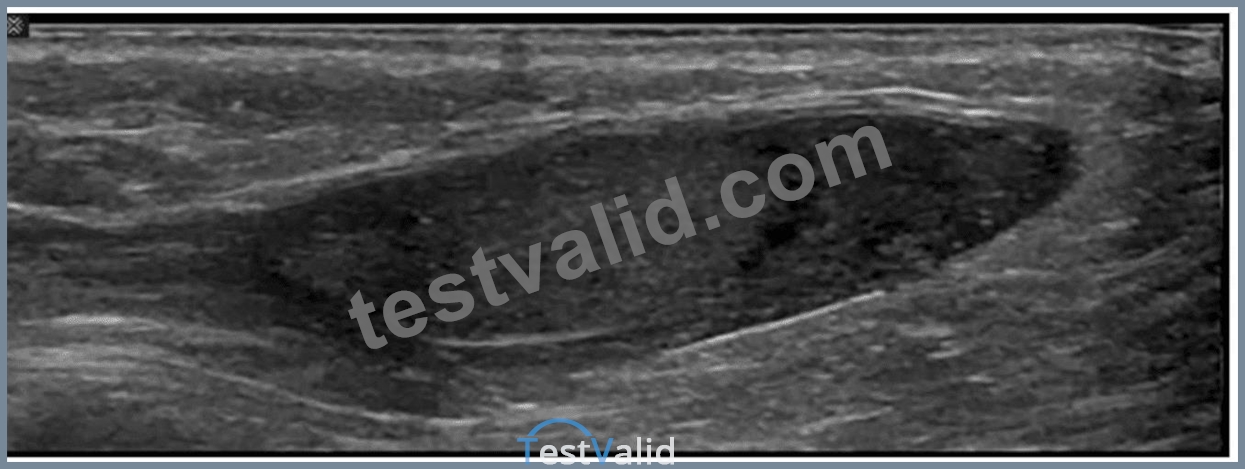
- A. Pyocele
- B. Bell clapper deformity
- C. Inguinal hernia
- D. Cryptorchidism
Answer: D
Explanation:
The ultrasound image shows an ovoid, homogeneously hypoechoic soft tissue structure located in the inguinal canal, surrounded by echogenic fat and soft tissue. This is consistent with an undescended testis, also known as cryptorchidism.
Cryptorchidism refers to the failure of one or both testes to descend into the scrotal sac. On ultrasound, the undescended testis typically appears:
* Ovoid in shape
* Homogeneous and hypoechoic compared to scrotal testis
* Located in the inguinal canal or, less commonly, within the abdomen
* Smaller in size than a normally descended testis
Comparison of answer choices:
* A. Bell clapper deformity refers to an anatomic predisposition for testicular torsion where the tunica vaginalis surrounds the entire testis and epididymis-usually a clinical rather than directly sonographic diagnosis.
* B. Inguinal hernia appears as bowel or omentum within the inguinal canal or scrotum with peristalsis or fat-no bowel loops are seen here.
* C. Pyocele is a complex fluid collection around the testis (usually with septations and internal echoes)- not evident in this image.
* D. Cryptorchidism - Correct. The findings match those of an undescended testis in the inguinal canal.
References:
Rumack CM, Wilson SR, Charboneau JW, Levine D. Diagnostic Ultrasound, 5th ed. Elsevier; 2017.
Dogra VS, Gottlieb RH, Rubens DJ, Oka M. Sonography of the scrotum. Radiology. 2003;227(1):18-36.
AIUM Practice Parameter for the Performance of Scrotal Ultrasound Examinations (2021).
NEW QUESTION # 84
Which condition is most consistent with the findings in the image below?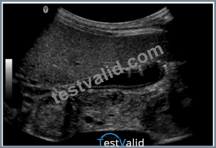
- A. Adenomyomatosis
- B. Gangrenous cholecystitis
- C. Emphysematous cholecystitis
- D. Porcelain gallbladder
Answer: C
Explanation:
The ultrasound image shows echogenic foci with dirty shadowing and reverberation artifacts within the gallbladder wall and lumen. These features are characteristic of emphysematous cholecystitis, a severe, life- threatening variant of acute cholecystitis caused by gas-forming organisms (e.g., Clostridium or E. coli) infecting the gallbladder wall.
Sonographic features of emphysematous cholecystitis:
* Echogenic gas within the gallbladder wall or lumen
* Reverberation or "dirty" shadowing artifacts
* May show intramural gas bubbles or "ring-down" artifact
* Often seen in diabetic or immunocompromised patients
* No gallstones may be present ("acalculous cholecystitis")
Clinical context:
* More common in elderly men and diabetics
* Presents with right upper quadrant pain, fever, and leukocytosis
* Surgical emergency due to risk of perforation and sepsis
Differentiation from other options:
* A. Adenomyomatosis: Involves gallbladder wall thickening with "comet tail" artifacts due to Rokitansky-Aschoff sinuses, not intramural gas.
* B. Porcelain gallbladder: Shows curvilinear calcification of the gallbladder wall - dense echogenic rim with posterior shadowing.
* C. Gangrenous cholecystitis: May show wall irregularity, intraluminal membranes, and absence of Doppler flow but lacks intramural gas.
References:
Rumack CM, Wilson SR, Charboneau JW, Levine D. Diagnostic Ultrasound. 5th Edition. Elsevier, 2018.
Chapter: Gallbladder and Biliary System, pp. 155-160.
American College of Radiology (ACR). Appropriateness Criteria for Right Upper Quadrant Pain, 2022.
Radiopaedia.org. Emphysematous cholecystitis: https://radiopaedia.org/articles/emphysematous-cholecystitis
NEW QUESTION # 85
Which technique would best assist the sonographer to verify the finding in this image obtained from the right upper quadrant?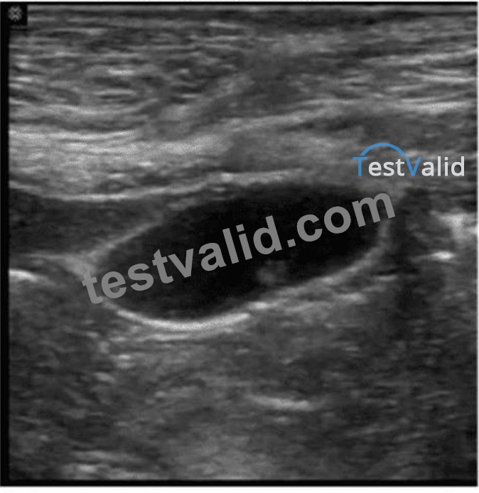
- A. Change the patient's position
- B. Assess for sonographic Murphy sign
- C. Use compound imaging
- D. Ask patient to perform Valsalva
Answer: B
Explanation:
The image demonstrates a gallbladder with a possible echogenic focus (likely a gallstone) and posterior acoustic shadowing. This is suggestive of cholelithiasis. To differentiate uncomplicated gallstones from acute cholecystitis, the most effective technique is to assess for a positive sonographic Murphy sign.
A positive sonographic Murphy sign refers to the presence of focal tenderness over the gallbladder when it is directly palpated with the ultrasound transducer. It is a strong indicator of acute cholecystitis when combined with other features such as gallbladder wall thickening, pericholecystic fluid, and gallstones.
Sonographic Murphy sign - key points:
* Assessed during real-time scanning
* Localized tenderness when pressure is applied over the gallbladder
* Highly sensitive for acute cholecystitis (especially in the presence of stones) Differentiation from other options:
* A. Use compound imaging: Improves image quality by reducing artifacts but does not verify tenderness or confirm acute inflammation.
* B. Change the patient's position: Helpful to confirm mobility of gallstones, but not diagnostic of inflammation.
* C. Ask patient to perform Valsalva: Used primarily in vascular studies (e.g., assessing for varicocele or venous reflux), not relevant here.
References:
Rumack CM, Wilson SR, Charboneau JW, Levine D. Diagnostic Ultrasound. 5th Edition. Elsevier, 2018.
Chapter: Gallbladder and Biliary System, pp. 148-152.
AIUM Practice Parameter for the Performance of an Ultrasound Examination of the Abdomen and/or Retroperitoneum, 2020.
Radiopaedia.org. Sonographic Murphy sign:https://radiopaedia.org/articles/sonographic-murphy-sign
NEW QUESTION # 86
Which technique is best for demonstrating the characteristic of the small hepatic lesion identified by the arrow on this image?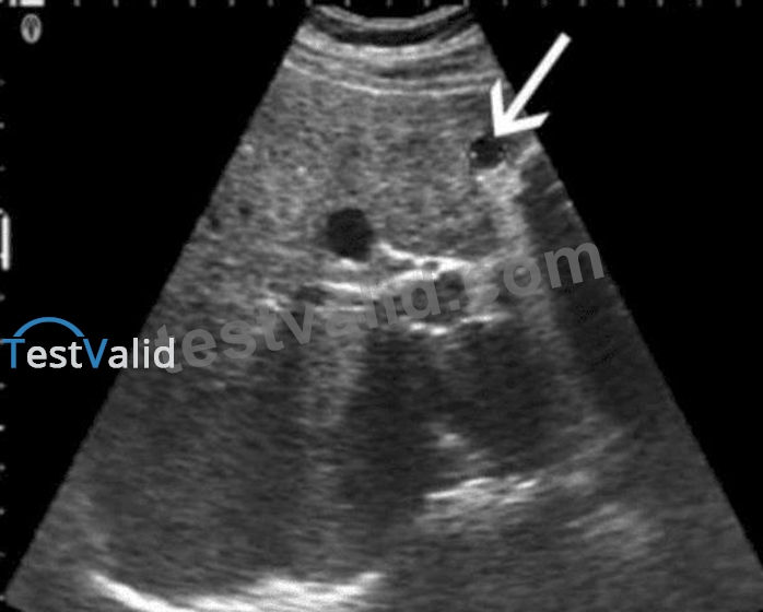
- A. Scan in upright position
- B. Use a standoff pad
- C. Decrease depth
- D. Move the transducer focus
Answer: B
Explanation:
The image shows a small hepatic lesion located very close to the anterior liver capsule, as indicated by the arrow. When imaging very superficial or near-field structures like subcapsular hepatic lesions, using a standoff pad is the most effective technique for optimizing visualization.
A standoff pad (also known as an acoustic stand-off or gel pad) helps increase the distance between the transducer and the superficial target. This improves the focus and beam shape for near-field imaging and minimizes reverberation and ring-down artifacts. It allows better evaluation of superficial lesions by positioning them within the focal zone of the transducer, which is usually set a few millimeters below the probe surface.
Differentiation from other options:
* A. Decrease depth: While reducing depth can help center deeper lesions in the field of view, it does not address issues with near-field resolution.
* B. Scan in upright position: This may help in gallbladder or fluid positioning but is not optimal for improving visualization of superficial liver lesions.
* C. Move the transducer focus: Adjusting focus deeper into the image won't enhance resolution of very superficial structures unless a standoff is used to bring the lesion into the focal zone.
References:
Rumack CM, Wilson SR, Charboneau JW, Levine D. Diagnostic Ultrasound. 5th Edition. Elsevier, 2018.
Chapter: Liver, pp. 80-84.
Kremkau FW. Sonography: Principles and Instruments. 9th Edition. Elsevier, 2015. Chapter: Image Formation and Optimization, pp. 114-117.
AIUM Practice Parameter for the Performance of an Ultrasound Examination of the Abdomen and/or Retroperitoneum, 2020.
NEW QUESTION # 87
Which condition puts the patient at greatest risk for a hematoma as a result of biopsy?
- A. Liver disease
- B. Acute renal failure
- C. Infection
- D. Hypertension
Answer: A
Explanation:
Patients with liver disease often have coagulopathy due to impaired synthesis of clotting factors. This places them at greater risk for bleeding or hematoma formation after biopsy. While hypertension may increase bleeding risk slightly, liver disease presents a significantly higher risk due to impaired coagulation.
According to the Society of Interventional Radiology (SIR) guidelines:
"Liver dysfunction is a significant risk factor for post-biopsy hemorrhage due to associated coagulopathy." Reference:
SIR Consensus Guidelines for Coagulation Parameters in Image-Guided Procedures, 2019.
American Association for the Study of Liver Diseases (AASLD), Practice Guidance, 2021.
-
NEW QUESTION # 88
Which scanning approach was utilized to obtain this image?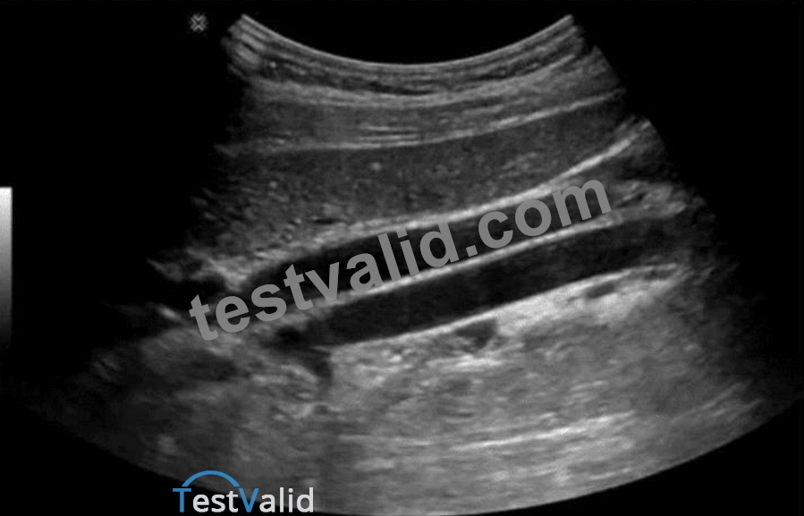
- A. Anterior
- B. Posterior
- C. Right coronal
- D. Left coronal
Answer: C
Explanation:
The ultrasound image provided shows the liver and diaphragm imaged in a coronal plane with characteristic rib shadows and costophrenic angles. The orientation of the image and the structures visualized suggest that the transducer is placed in the right mid-axillary line, with the sound beam directed coronally - this is a classic right coronal scanning approach.
Key features supporting this:
* The liver appears superiorly in the image.
* Multiple echogenic lines (representing the ribs) run obliquely, casting acoustic shadows.
* The diaphragm and adjacent lung base are seen clearly, which is commonly imaged through the right intercostal spaces in a coronal plane.
Comparison of answer choices:
* A. Anterior: Would show a more transverse view of the liver and not typically image the diaphragm and lung this way.
* B. Posterior: Not used for upper abdominal scanning due to shadowing from the spine and posterior ribs.
* C. Left coronal: Would show the spleen and left kidney - not the liver as seen here.
* D. Right coronal - Correct. This image was obtained using the right coronal (intercostal) approach through the right flank.
References:
Rumack CM, Wilson SR, Charboneau JW, Levine D. Diagnostic Ultrasound, 5th ed. Elsevier; 2017.
Hagen-Ansert SL. Textbook of Diagnostic Sonography, 8th ed. Elsevier; 2017.
AIUM Practice Parameter for the Performance of an Ultrasound Examination of the Abdomen and/or Retroperitoneum (2020).
NEW QUESTION # 89
Which liver neoplasm is associated with use of oral contraceptives and is most often seen in women under the age of 40?
- A. Adenoma
- B. Cavernous hemangioma
- C. Hepatoblastoma
- D. Hepatoma
Answer: A
Explanation:
Hepatic adenomas are benign liver tumors strongly associated with long-term use of oral contraceptives and are most frequently found in women under 40. Hepatoblastoma is seen in children; hepatoma (HCC) is a malignant tumor typically found in cirrhotic livers. Cavernous hemangioma is unrelated to oral contraceptives.
According to Rumack's Diagnostic Ultrasound:
"Hepatic adenomas occur predominantly in young women with a history of oral contraceptive use." Reference:
Rumack CM, Wilson SR, Charboneau JW, Levine D. Diagnostic Ultrasound. 5th ed. Elsevier, 2017.
WHO Classification of Tumours of the Digestive System, 5th ed., IARC, 2019.
-
NEW QUESTION # 90
Where is the most common location for a branchial cyst in relation to the thyroid?
- A. Anterior
- B. Lateral
- C. Posterior
- D. Medial
Answer: B
Explanation:
Branchial cleft cysts are congenital epithelial cysts that typically occur laterally in the neck, often anterior to the sternocleidomastoid muscle, and lateral to the thyroid gland. The second branchial cleft cyst is the most common type and is found in the lateral neck region.
* Medial (B) would be more consistent with thyroglossal duct cysts.
* Anterior (C) or posterior (D) do not specifically describe branchial cyst location relative to the thyroid.
Reference Extracts:
* Som PM, Curtin HD. Head and Neck Imaging. 5th ed. Elsevier, 2011.
* Rumack CM, Wilson SR, Charboneau JW, Levine D. Diagnostic Ultrasound. 5th ed. Elsevier, 2017.
-
NEW QUESTION # 91
Which structure is indicated by the arrow in this image?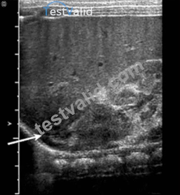
- A. Adrenal gland
- B. Kidney
- C. Diaphragm
- D. Bowel loop
Answer: A
Explanation:
The structure indicated by the arrow in the ultrasound image is the adrenal gland. On ultrasound, the adrenal gland in neonates and infants is relatively large and has a distinctive "Y" or "V" shape in the transverse view.
It is located superior and slightly medial to the upper pole of the kidney.
In this image, the arrow is pointing to a hypoechoic, curved structure with a thin echogenic central stripe, representing the fetal adrenal gland. This echogenic stripe corresponds to the adrenal medulla, while the surrounding hypoechoic area represents the cortex.
Differential features:
* A. Kidney: While the kidney is visualized posterior to the adrenal gland and shows a reniform shape with a central echogenic sinus and peripheral cortex, it is not the structure being directly pointed to by the arrow.
* B. Bowel loop: Bowel has variable echogenicity with peristalsis and shadowing from air. It does not have the consistent morphology or location seen in the image.
* C. Diaphragm: Appears as a thin, hyperechoic linear structure separating the thoracic cavity from the abdomen. It is seen more superiorly than the indicated structure and lacks the "Y" or "V" adrenal configuration.
Key Anatomical Landmarks:
* The adrenal glands are located in the retroperitoneum, superior to the kidneys, and appear prominent on ultrasound in neonates.
* In transverse view, the right adrenal gland is anterior to the crus of the diaphragm and posterior to the inferior vena cava (IVC).
References:
Rumack CM, Wilson SR, Charboneau JW, Levine D. Diagnostic Ultrasound. 5th Edition. Elsevier, 2018.
Chapter: Adrenal Glands and Retroperitoneum, pp. 291-295.
American Institute of Ultrasound in Medicine (AIUM) Practice Parameter for the Performance of an Ultrasound Examination of the Abdomen and/or Retroperitoneum. 2020.
NEW QUESTION # 92
Which vascular condition is most consistent with patent cutaneous para-umbilical channels and portal hypertension?
- A. Splenic vein varices
- B. Coronary vein varices
- C. Esophageal varices
- D. Caput medusae
Answer: D
Explanation:
Caput medusae refers to dilated paraumbilical veins due to portal hypertension. When portal venous pressure rises, collateral channels may open along the ligamentum teres and recanalized paraumbilical vein, resulting in visible dilated veins radiating from the umbilicus.
* Esophageal varices (B) are gastroesophageal collaterals.
* Coronary vein varices (C) involve gastric veins.
* Splenic vein varices (D) are typically localized to the splenic hilum.
Reference Extracts:
* Rumack CM, Wilson SR, Charboneau JW, Levine D. Diagnostic Ultrasound. 5th ed. Elsevier, 2017.
* Gore RM, Levine MS. Textbook of Gastrointestinal Radiology. 4th ed. Saunders, 2015.
-
NEW QUESTION # 93
Which sonographic finding is commonly associated with transitional cell cancer of urinary bladder?
- A. Polypoidal non-mobile focal mass
- B. Flat sessile lesion
- C. Diffuse wall thickening
- D. Ulcerated solid infiltrative lesion
Answer: A
Explanation:
Transitional cell carcinoma (TCC) typically presents as a non-mobile, polypoidal, focal intraluminal mass projecting from the bladder wall. Mobility of the lesion helps differentiate TCC from blood clots or debris.
According to Rumack's Diagnostic Ultrasound:
"Bladder TCC most often appears as a non-mobile, polypoid mass attached to the bladder wall." Reference:
Rumack CM, Wilson SR, Charboneau JW, Levine D. Diagnostic Ultrasound. 5th ed. Elsevier, 2017.
AIUM Practice Parameter for Bladder Ultrasound, 2020.
-
NEW QUESTION # 94
Which of the following is a possible early complication of a renal transplant?
- A. Acute tubular necrosis
- B. Ureterocele
- C. Transitional cell carcinoma
- D. Transplant artery stenosis
Answer: A
Explanation:
Acute tubular necrosis (ATN) is the most common cause of early graft dysfunction following renal transplantation. It results from ischemia-reperfusion injury during the transplantation process. Ultrasound findings may be nonspecific but Doppler may show elevated resistive indices.
Ureterocele (A) is a congenital anomaly.
Transplant artery stenosis (C) is a late complication.
Transitional cell carcinoma (D) is rare and not typically an early complication.
Reference Extracts:
Middleton WD, Kurtz AB, Hertzberg BS. Ultrasound: The Requisites. 3rd ed. Elsevier, 2015.
Rumack CM, Wilson SR, Charboneau JW, Levine D. Diagnostic Ultrasound. 5th ed. Elsevier, 2017.
-
NEW QUESTION # 95
Which condition is characterized by abnormal dilatation of veins of the pampiniform plexus and most commonly affects the left testicle?
- A. Hematocele
- B. Hydrocele
- C. Spermatocele
- D. Varicocele
Answer: D
Explanation:
A varicocele is an abnormal dilatation of the pampiniform plexus veins, usually seen on the left side due to the perpendicular insertion of the left testicular vein into the left renal vein, making it more susceptible to elevated venous pressure. Sonographically, varicoceles appear as multiple serpiginous anechoic tubular structures that show venous flow on color Doppler, often accentuated with Valsalva maneuver.
Hydrocele (A) is a fluid collection surrounding the testis.
Hematocele (C) is blood within the tunica vaginalis.
Spermatocele (D) is a cystic lesion arising from the epididymis.
Reference Extracts:
Dogra VS, Bhatt S. "Sonographic evaluation of testicular varicoceles." Journal of Ultrasound in Medicine.
2004;23(6): 829-838.
Rumack CM, Wilson SR, Charboneau JW, Levine D. Diagnostic Ultrasound. 5th ed. Elsevier, 2017.
-
NEW QUESTION # 96
Which portion of the biliary system is last to become dilated with biliary obstruction at the ampulla of Vater?
- A. Cystic duct
- B. Common bile duct
- C. Common hepatic duct
- D. Peripheral bile ducts
Answer: D
Explanation:
In biliary obstruction (such as at the ampulla of Vater), dilation begins proximally and progresses peripherally. The intrahepatic peripheral bile ducts are the last to dilate because backpressure takes time to propagate. Early dilation is typically seen in the common bile duct and common hepatic duct.
According to Rumack's Diagnostic Ultrasound:
"The intrahepatic peripheral bile ducts dilate last in the setting of progressive biliary obstruction." Reference:
Rumack CM, Wilson SR, Charboneau JW, Levine D. Diagnostic Ultrasound. 5th ed. Elsevier, 2017.
AIUM Practice Parameter for the Performance of Abdominal Ultrasound, 2020.
-
NEW QUESTION # 97
Which disease process may cause numerous shadowing calcifications to form within the spleen?
- A. Histoplasmosis
- B. Sickle cell anemia
- C. Thalassemia
- D. Non-Hodgkin lymphoma
Answer: A
Explanation:
Histoplasmosis is a fungal infection that can lead to granulomatous disease. Chronic granulomatous infections may result in multiple splenic calcifications that appear as small echogenic foci with shadowing on ultrasound. Other infectious granulomas (e.g., tuberculosis) may present similarly.
According to Rumack's Diagnostic Ultrasound:
"Granulomatous infections such as histoplasmosis and tuberculosis may produce multiple splenic calcifications, often with shadowing." Reference:
Rumack CM, Wilson SR, Charboneau JW, Levine D. Diagnostic Ultrasound. 5th ed. Elsevier, 2017.
AIUM Practice Parameter for the Performance of Abdominal Ultrasound Examinations, 2020.
-
NEW QUESTION # 98
Which finding is most likely demonstrated in this image?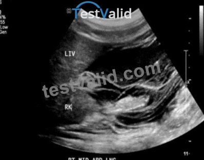
- A. Bowel obstruction
- B. Ascites
- C. Hydropic gallbladder
- D. Hemoperitoneum
Answer: B
Explanation:
The ultrasound image shows an anechoic (black) fluid collection in the perihepatic and perirenal spaces. The fluid outlines the liver (LIV) and right kidney (RK), which is characteristic of free fluid in the peritoneal cavity - consistent with ascites.
Sonographic features of ascites:
* Anechoic (or hypoechoic) fluid in dependent areas of the abdomen
* Seen surrounding the liver, spleen, and intestines
* Can be free-flowing or loculated
* Bowel loops may be floating or displaced centrally
This image is consistent with a typical finding of ascites: free fluid in Morison's pouch (hepatorenal recess), a common site for fluid accumulation.
Differentiation from other options:
* A. Hydropic gallbladder: Refers to an enlarged gallbladder filled with clear bile; not visible in this image.
* B. Hemoperitoneum: May appear similar to ascites, but usually has complex echogenicity or layering if acute; clinical context (trauma, bleeding) is essential for diagnosis.
* C. Bowel obstruction: Would show dilated, fluid-filled bowel loops with peristalsis or to-and-fro motion, not evident here.
References:
Rumack CM, Wilson SR, Charboneau JW, Levine D. Diagnostic Ultrasound. 5th Edition. Elsevier, 2018.
Chapter: Peritoneal Cavity and Abdominal Trauma, pp. 125-130.
American Institute of Ultrasound in Medicine (AIUM). Practice Parameter for the Performance of a Focused Assessment with Sonography for Trauma (FAST) Examination, 2020.
Radiopaedia.org. Ascites (ultrasound): https://radiopaedia.org/articles/ascites-ultrasound
NEW QUESTION # 99
......
Ace AB-Abdomen Certification with 165 Actual Questions: https://www.testvalid.com/AB-Abdomen-exam-collection.html
PASS ARDMS AB-Abdomen EXAM WITH UPDATED DUMPS: https://drive.google.com/open?id=1aFIBVcPCHyIkSD5360_1AKVWq2STYCPz